A Journey Toward Normal Vision
I was born with what most would describe as a very "complex visual system". A variety of organic problems conspired to insure that my vision, even with correction, was never ideal. For almost all of my life I have worn corrective lenses. The lenses goal was to alleviate widely varying levels of visual acuity in both eyes, as well as a dramatic muscle imbalance and dominant eye problem. Over four decades, my eyes became more disparate and my depth perception, which was minimal, became almost non-existent. I had always wanted to know what it was like to see normally, but was a little nervous about surgical correction. Well, everyone hits a wall and I hit mine. I had an opportunity to minimize the risk of the surgeries by taking some serious time off of my work life and decided to go for it. So that you can understand what was involved, I'll begin at the beginning.
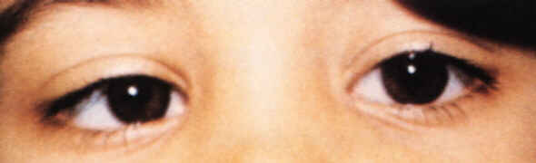
To begin with, I have always had some pretty severe muscle problems in my eyes. Some call this "lazy eye". However, my condition went beyond just lazy eye. The first part of the muscle problem is called accommodative esotrpoia and looks like this:
Notice how one eye appears to be turning in? In my case, one eye would turn in by itself and both would turn in severely the more closely I tried to focus on a near object (ex: reading). In addition to this problem, my left eye had an up and down muscle problem when I tried to focus at an object that was above and to the side of my direct line of sight. My right eye was the one that turned in the most, but my left eye was dominant, even though it was very near-sighted. Now is probably a good time to describe my visual acuity. My right eye (the one that really turned in) was slightly far-sighted with a good deal of astigmatism. My left eye was very near-sighted and also had a good deal of astigmatism. In order to try to hold my eyes relatively straight, prisms were added to my lenses to "re-direct" the image path for my eyes. Also, progressive bifocals were used in order to try and minimize the accommodative issues associated with close up work. The bottom part of my lenses were the equivalent of a +2.5 pair of reading glasses. With all of this going on, the lens shape and thickness made contacts not possible. Moreover, my dominant eye was causing a ton of problems. Essentially, even though I could see out of my right eye, my brain pretty much ignored the input. That was hell on my depth perception. For all intents and purposes, my brain just "inferred" depth, or 3D, and I just went on my merry way. How bad was my depth perception? Pretty rotten.
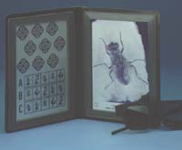
What you see above is one of the classic Randot test kits used to measure depth of field sensitivity. You put on these special glasses and stare at the images. Normal eyes would see the images stereoscopically when using the test glasses. For instance, the fly's wings would appear to have depth and look like they were coming off the page. At the top left you can see nine diamond shaped images. Each of those diamonds has a part that should appear to have depth during the test. Going left to right and then down each row, the amount of depth gets reduced. A normal pair of eyes should get to, on average, the sixth image. I could only get the first one right.
So my depth perception stunk, my eyes still crossed, and my visual acuity was pretty unbalanced between my two eyes. I really couldn't even make it across a room at night without my glasses. And I had to "sneak up" on things to pick them up off of a table by sliding my hand along the table to "triangulate" the item's position. Without glasses, life stunk. With them, things were marginally better. I decided that it was time to try to make my life a little better. I started working with Dr. Samuel Yankelove, in Houston, to put together a game plan for addressing my issues. Dr. Yankelove had performed LASIK on my wife and she was very pleased with the results, so I already had a great deal of trust invested with this board certified refractive surgeon. We agreed on a course of action that would require a combination of LASIK correction in both eyes and then corrective surgery for my muscles. I easily qualified as a LASIK candidate. My pupils were not too large and my corneas were not too thin. LASIK requires a bit of suction to create a flap. The muscle surgery would require that the surface of my eyes be disrupted, which would cause difficulty in creating that suction until fully healed. It was decided to do LASIK first and then the muscles.
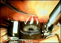
In order to shape the cornea with a laser, it must be exposed. My doctor used a device called a micro-keratome to attach a cutter to the eye (with suction) and then make a flap with that cutter. Some doctors now use a laser to cut this flap. This procedure is done while you are awake. However, your eyes have been given a lot of numbing drops and you can't really blink because your eyelids are being held open with retractors. Believe it or not, this doesn't really hurt.
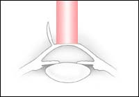
Once the flap is open, everything is blurry, but you are asked to focus on a red light overhead. The doctor feeds in the parameters and the laser quickly re-shapes your cornea. There is a slight aroma of burning hair. It's a little disquieting if you aren't aware that it is coming! The flap is smoothed back into place and irrigated. Some pure oxygen is gently blown around the edges of the flap and you are done. It is usually a bloodless procedure. The fact that you most likely have a Valium in your body makes things even easier. Here's an older video of a LASIK procedure if you are interested (big file).
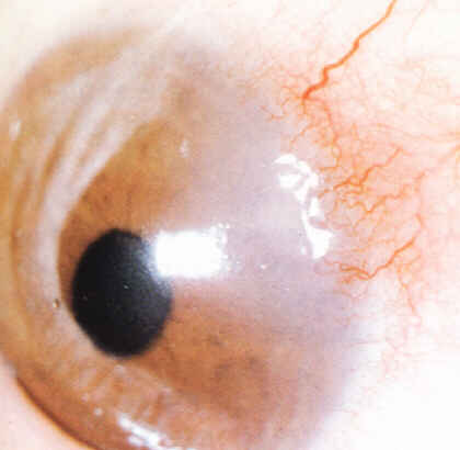
Remember how I said that LASIK is usually a bloodless procedure? Sometimes there is a little bleeding. It is almost always nothing to worry about. What you see above is a close-up picture of an eye that has a condition called pannus. My eyes have pannus. This is a condition where tiny blood vessels have crept onto the cornea. There are varying degrees of severity that can occur that range from what you see here, vessels at the edge of the cornea, to much more dramatic infiltration. My eyes exhibited the mild pannus variety. When the flaps were cut, there was a little bleeding. It was easily addressed and was no cause for concern. It was time for me to heal up from LASIK and prepare for the muscle surgery.
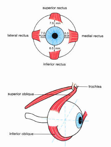
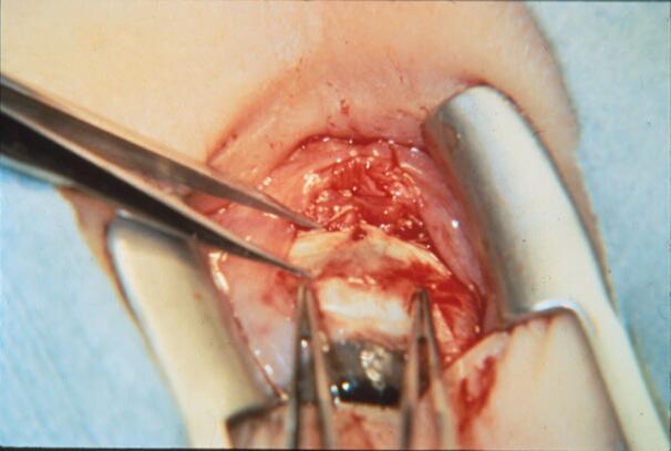
There are six muscles that control the movement of each eye ball. The image above shows the six muscles of the right eye. My muscle surgeries were done on both eyes under general anesthetic by Dr. James Lai, of Houston Eye Associates. The interior muscles of both eyes were detached from the "globe", shortened, and re-attached, with dissolvable stitches, onto new areas. The reduction of tension was the equivalent of about twenty eight diopters of prism correction. In addition, the muscle that controls the up and down movement of my left eye had a similar procedure performed. Now, if you think that you have a strong constitution and can handle a photo of what the actual muscle surgery looks like, click here. The forceps (up and down) show where the muscle used to be attached, the calipers (left to right angle) measure the distance to the new area where the muscle (the red blob with the white thread (stitches) will be attached. The whole procedure, including coming out of anesthesia, took a little longer than 3 hours. Within 48 hours of my procedure, I was seeing six out of nine on the Randot test. Normal stereoscopic vision. And my eyes were straight!
Now, it is not completely uncommon for a person to need a touch-up, or "enhancement" to their LASIK procedure. For a variety of reasons, including healing and others, the re-shaping of the cornea may not provide the desired level of correction. Usually, though, the surgeon waits many months to allow for complete healing to determine if an enhancement is actually necessary. An enhancement is usually a simple procedure that does not require the cutting of a new flap. Why? The top layer of the cornea, called the epithelium, is constantly regenerating itself. The flap surface from the original cut is completely healed in a few weeks, but the "interior" part of the flap can actually take many years to fully heal. To perform an enhancement, the surgeon numbs the eye and retracts the eyelids, just like in the original procedure. He/she then uses a special tool to gently separate the epithelial cells over the flap and then carefully re-open the original flap. Dr. Yankelove describes the "seal" as being like Velcro: pretty strong, but still easy to open. I needed an enhancement to my right eye.
Like I said, an enhancement is usually pretty easy. Well, I became a first for Dr. Yankelove. I went in to have the enhancement performed and he couldn't get the flap opened up. That was a first for him. Remember how I described the eye condition known as pannus? Those tiny little blood vessels had crept back onto my cornea after being severed. They created a reinforced structure across the flap margin that made it very difficult for the doctor to get the flap started. He kept trying, but all that wound up happening was that the epithelial cells over the flap just kept on moving around and getting abraded. Dr. Yankelove decided to just stop because he wasn't getting anywhere with the flap and he didn't want to cause any more corneal abrasion. He put a "bandage contact" on my eye and told me to come see him the next day in his office so that we could put together a game plan.
I showed up as instructed and got a thorough examination. I had a little abrasion from trying to get the flap open and I had also developed a very mild case of a condition called diffuse lamellar keratitis (DLK). It is a sterile inflammation that can get pretty bad if not treated. My DLK looked something like this diagram:
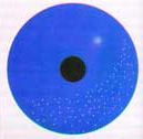
The DLK are those little white dots. So we get the corneal abrasion taken care of and the DLK cleared up. I wear the contact for about a week and then I have to wait two to three weeks before we can re-attempt the enhancement. The rule is that you have to wait at least two weeks after you've worn a contact before you can have the laser work on you. I return to the surgical center for a second try at my enhancement. Dr. Y finds a clear spot to get the flap started and is successful. It takes a very aggressive approach to get the whole flap to come up and he winds up having to gently cut a lot of those pannus blood vessels. But the flap does completely open and I received my enhancement! The follow up visit the next day showed that the DLK had returned, but this was expected and is treated the in the same manner as before.
The net of all of this is that I can now see in 3D. I'm about 20/25 in my left eye and will most likely be 20/20, or better, in my right eye. I don't wear corrective lenses anymore, except for long sessions of reading (hey, I'm over 40). I've given myself the gift of vision... with a lot of help from to very special doctors.